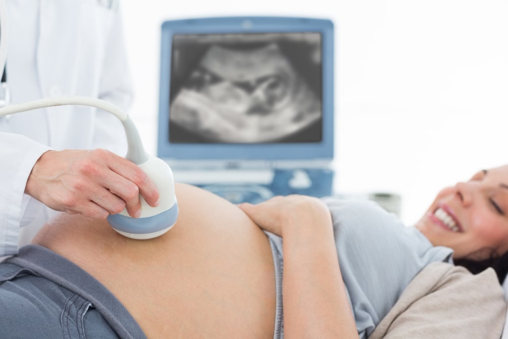
Ultrasound devices; It works with the principle that high-frequency sound waves emitted from the end of the device touching the mother are reflected from tissues of different densities and the image formed is reflected on a screen.
In traditional two-dimensional ultrasound, the reflections come straight, whereas in four-dimensional ultrasound, tissues can be imaged in detail because they come from different angles.
4D ultrasound ensures that the excitement of pregnancy turns into beautiful memories as families wonder who and what their baby looks like the most after their health condition.
Detailed ultrasound examination during pregnancy, 18-24 weeks of pregnancy. It is carried out in weeks. If an evaluation cannot be made due to the baby's position or if a suspicious finding is found, the procedure is repeated within a week.
The content of our website is prepared to inform visitors. The information provided on the site can never replace the advice or consultation of a physician.
© 2026 All Digital Activities are managed by Deniz-Web Agency.
